Detecting possible eye conditions or eye changes that may affect the clarity, comfort or health of your eyes should be the motivation for adhering to annual visual examinations for us all. Early detection of possible visual system problems may avoid loss of function or vision.
As an independent practice, we are able to allocate one hour of working time to each patient as standard. This allows for comprehensive evaluation and assists in better decision making, which in turn benefits the patient and helps attain the service level we strive for.
THE VISUAL EVALUATION MAY INCLUDE SOME IF NOT ALL OR MORE OF THE FOLLOWING:
- Comprehensive case history
- Taking and recording visual acuity with and without spectacles/contact lenses
- Auto and balanced subjective refraction
- Binocular muscle balances, accommodation, depth perception and colour vision
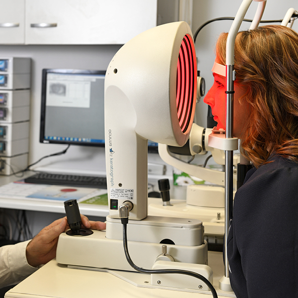
- The external eye – Slit lamp microscope – Corneal topography – Digital Dry Eye evaluation
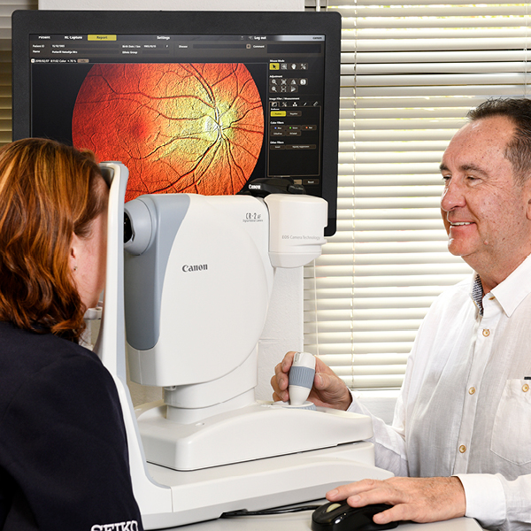
- The internal eye – Slit lamp microscope – Ophthalmoscopy – Retinal photography (24 megapixels)
- Non-contact tonometry – Intraocular pressures (Puff Glaucoma screening for Discovery Vitality points)
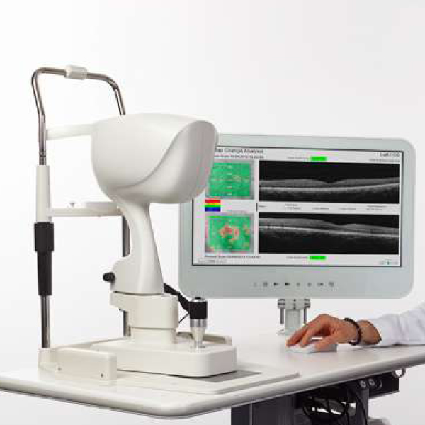
- OCT – Accurate digital screening for glaucoma – Cornea – Retina – Advanced contact lens fitting
EQUIPMENT USED DURING THE EXAMINATION ARE NON-INVASIVE AND MAY INCLUDE THE FOLLOWING:
Our Ophthalmoscope is a hand held illuminated variable focus instrument, used to view the inner eye from close to the patient’s pupil.
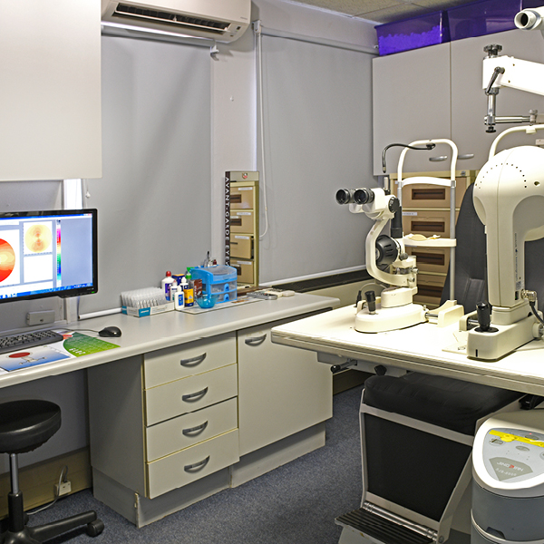
Our Slit lamp microscope is an instrument used by eye care practitioners to specifically view and evaluate the anterior eye structures. These would include from the outer tear layer, through the cornea, anterior chamber, iris, lens and vitreous of the eye. It is also extensively used in fitting contact lenses to ensure an accurate fit and assess correct physiological responses.